

Introduction
Before you ever opened your eyes to see the world, a silent chemical process was already deciding how clearly you’d see it. New research shows that vitamin A vision development plays a starring role in building sharp central vision, and the discovery is rewriting what scientists thought they knew about how the human eye forms.
Scientists at Johns Hopkins University have uncovered how the human eye develops sharp central vision before birth. Their findings could pave the way for new treatments to restore sight in people suffering from serious eye diseases like macular degeneration and glaucoma.
This isn’t just a lab curiosity. Millions of people lose their sharp vision every year due to age-related eye conditions that currently have no cure. Therefore, understanding vitamin A vision development could be the first step toward rebuilding damaged sight artificially. old muscle stem cells and other regenerative research.
The Hidden Chemistry Behind Vitamin A Vision Development
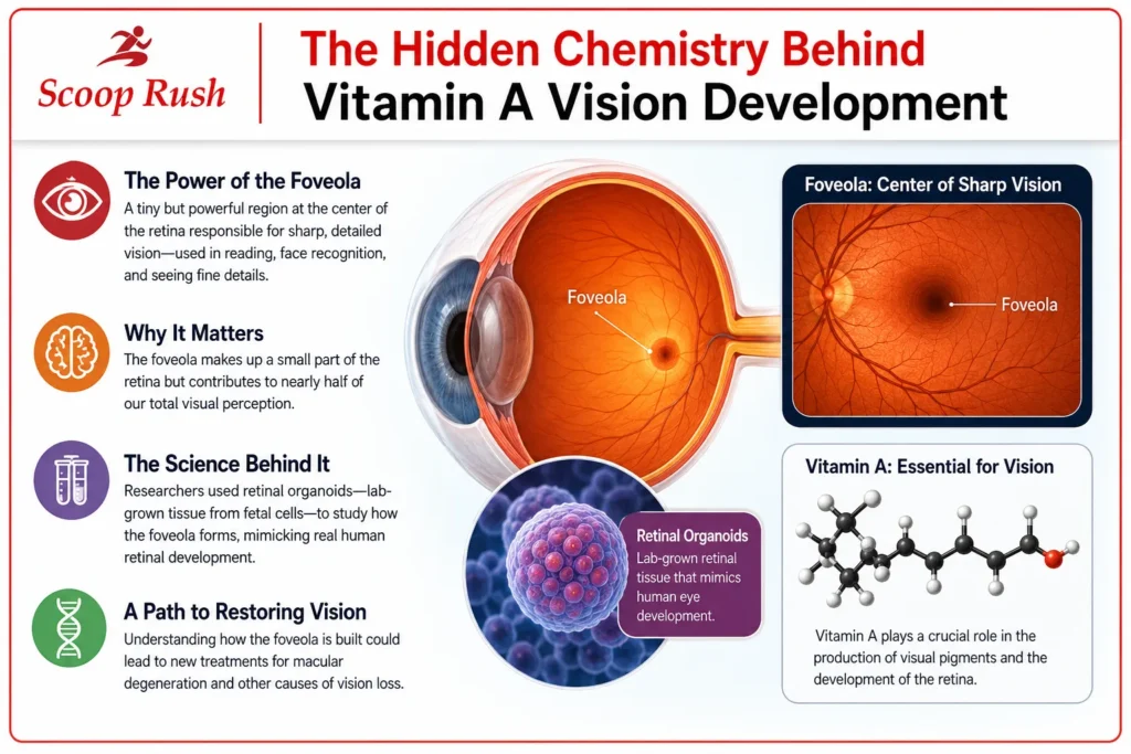
The study, published in the journal Proceedings of the National Academy of Sciences, focused on a tiny but powerful part of the eye called the foveola. This small region sits at the center of the retina and is responsible for your sharpest vision, the kind you use for reading, recognizing faces, or spotting details from far away.
Although the foveola makes up only a small fraction of the retina, it accounts for nearly half of a person’s total visual perception. That’s a massive amount of visual power packed into a tiny space.
To study how this region forms, researchers used retinal organoids. These are small pieces of retinal tissue grown in a lab from fetal cells. Because they closely mimic the structure and function of a real retina, organoids allow scientists to observe eye development in ways that would otherwise be impossible, similar to how researchers study neurons breaking DNA to build the brain to understand early development.
Why the Foveola Matters So Much
Robert J. Johnston Jr., associate professor of biology at Johns Hopkins and the study’s lead researcher, explained why this region deserves so much attention. He called it a key step toward understanding the center of the retina, since it’s often the first part of the eye damaged in people with macular degeneration.
In other words, if scientists can understand how the foveola is built, they may eventually learn how to rebuild it after disease damages it. That’s a big deal for anyone facing vision loss later in life, and it echoes a broader pattern in science where understanding how the brain visualizes things, such as in cases of aphantasia, continues to reshape what we know about perception.
The Cone Cells That Make Color Vision Possible
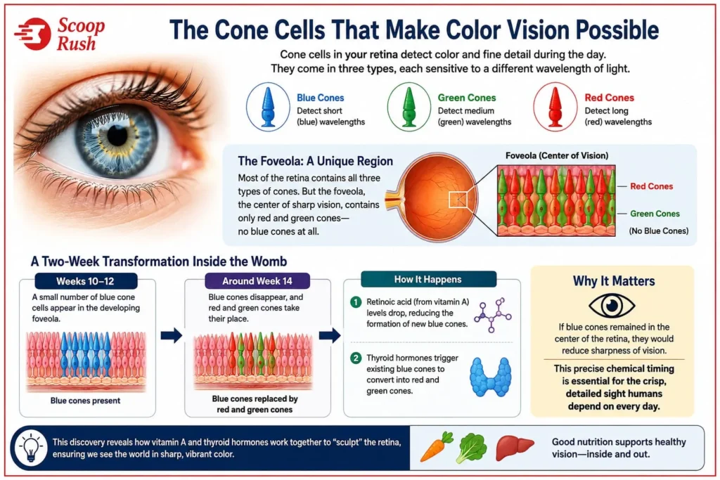
To understand this discovery, it helps to know a bit about cone photoreceptors. These are the specialized cells in your retina that allow you to see color and fine detail during the day. Cone cells develop into one of three types: blue, green, or red, and each type detects a different wavelength of light.
Most of the retina contains a mix of all three cone types. However, the foveola is different. It contains only red and green cones, with no blue cones at all. Scientists have long wondered how this unusual pattern comes together, and until now, nobody had a clear answer. Most of the retina contains a mix of all three cone types. However, the foveola is different. It contains only red and green cones, with no blue cones at all. Scientists have long wondered how this unusual pattern comes together, and until now, nobody had a clear answer for this part of vitamin A vision development.
A Two-Week Transformation Inside the Womb
The research team tracked exactly when and how this pattern forms. During weeks 10 through 12 of fetal development, a small number of blue cone cells appear in the developing foveola. Then, by around week 14, something surprising happens. Those blue cones disappear, and red and green cones take their place.
This transformation happens through two separate biological steps.
First, levels of retinoic acid, a molecule produced from vitamin A, begin to drop. This decrease reduces the formation of new blue cone cells. Second, thyroid hormones step in and trigger the remaining blue cones to convert into red and green cones.
Johnston emphasized why this sequence matters so much for vision quality. If blue cones remained in the center of the retina, they would reduce the sharpness of a person’s vision. As a result, this precise chemical timing is essential for building the crisp, detailed sight that humans depend on every day.
Rewriting Three Decades of Vision Science
Perhaps the most striking part of this discovery is how it overturns a theory that has guided vision research for about 30 years. Scientists previously believed that blue cone cells formed in the center of the retina and then physically moved outward, clearing space for red and green cones to take over.
However, this new study tells a different story. Instead of migrating away, blue cones actually stay in place. They simply change their identity, transforming directly into red and green cones over time.
This finding matters because it shifts the entire framework scientists use to understand retinal development. Additionally, it opens new questions about what else might trigger similar transformations in other parts of the body, in much the same way that a 3.2-billion-year-old enzyme recently forced scientists to rethink assumptions about how ancient biology worked. This finding matters because it shifts the entire framework scientists use to understand vitamin A vision development. Additionally, it opens new questions about what else might trigger similar transformations in other parts of the body.
Why This Discovery Took So Long
You might wonder why it took decades to figure this out. Part of the challenge lies in the animals typically used for vision research. Mice and fish, two of the most common lab animals, don’t develop the same three-cone arrangement found in human eyes.
Because of this, scientists lacked a good model to study the process directly. Human retinal organoids solved that problem. By growing lab-based tissue from fetal cells, researchers finally had a way to watch this transformation unfold step by step, a research approach not unlike how scientists identified a new beetle species by carefully observing traits that had gone unnoticed for years.
What This Means for Future Eye Treatments
This research isn’t just about satisfying scientific curiosity. It has real potential to shape future treatments for people living with vision loss.
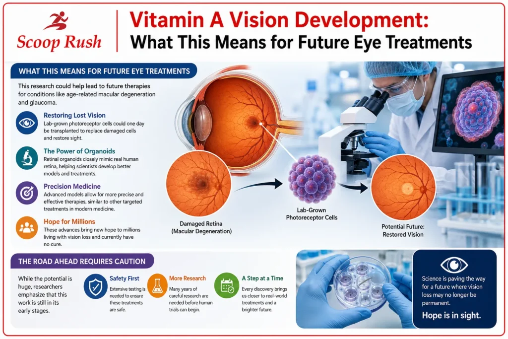
Age-related macular degeneration and glaucoma both damage the retina and can eventually lead to blindness. Currently, there is no cure for macular degeneration, which makes discoveries like this one especially valuable.
Researchers hope to use organoid technology to eventually create healthy, made-to-order photoreceptor cells. These lab-grown cells could potentially be transplanted into damaged eyes to restore lost vision. Meanwhile, scientists continue refining these organoids so they more closely match the function of an actual human retina, a strategy that mirrors ongoing work using silica nanoparticles for prostate cancer treatment to precisely target damaged cells.
Better models mean better cells, and better cells mean a stronger foundation for cell replacement therapy. Consequently, this research brings scientists one step closer to real-world treatments, even though clinical use is still years away.
The Road Ahead Requires Caution
Despite the excitement, researchers are quick to point out that this work is still in its early stages. Much more research is needed to confirm both the safety and effectiveness of these approaches before they can ever be tested in patients.
This kind of caution is standard in medical science, and for good reason. Treatments involving cell transplantation require extensive testing to ensure they work safely inside the human body. Still, every discovery like this lays another brick on the path toward future therapies.
A Bigger Picture: Vitamin A’s Long History in Vision Science
This discovery builds on a much longer story about vitamin A vision development and its role in eye health. Vitamin A is essential for life, yet the human body cannot produce it on its own. It must come from the diet, primarily through foods like carrots, leafy greens, and animal liver.
For centuries, people have understood that vitamin A deficiency harms vision. Night blindness caused by low vitamin A levels was documented as far back as 3500 BC, with ancient remedies even suggesting the consumption of liver, a food naturally rich in the vitamin.
Modern science eventually confirmed why this remedy worked. A modified form of vitamin A, known as retinal, combines with a protein called opsin to create the visual pigments found in photoreceptor cells. When light hits these pigments, it triggers the chemical reaction that allows the eye to detect and process images.
Without enough vitamin A, these visual pigments cannot form properly, and photoreceptors become less sensitive to light. This is why vitamin A deficiency causes night blindness before it causes more severe vision problems, a reminder of how deeply diet shapes long-term health, much like ongoing debates around the risks of a no-sugar diet.
Beyond basic vision, retinoic acid, the active form of vitamin A, has been shown to influence many other processes in the body. It affects the pace at which certain cells, especially photoreceptors, develop. Interestingly, it has also proven effective in treating a form of leukemia, transforming what was once one of the deadliest cancers into one with a remission rate above 90 percent when caught early. Nutritional factors influencing disease outcomes are a growing research theme, as seen in studies linking omega-3 fish oil supplements to brain health.
Key Takeaways
This new study reveals that vitamin A vision development involves far more than general eye health support. It actively shapes the precise arrangement of cone cells that gives humans their sharp central vision.
Through a coordinated process involving retinoic acid and thyroid hormones, blue cone cells transform into red and green cones during a critical window of fetal development. This finding overturns a 30-year-old theory that assumed these cells simply migrated out of the way.
As a result, scientists now have a clearer path toward understanding how the human retina builds its most vital region, the foveola. Ultimately, this knowledge could help researchers develop lab-grown retinal tissue capable of restoring vision lost to diseases like macular degeneration and glaucoma.
While treatments are still years away, this discovery marks meaningful progress toward a future where blindness caused by retinal damage may finally become treatable.
FAQs
How does vitamin A help build sharp vision before birth?
Vitamin A produces a molecule called retinoic acid, which helps reduce the formation of blue cone cells in the developing retina. This process, combined with thyroid hormone activity, helps shape the arrangement of cone cells needed for sharp central vision.
What is the foveola and why is it important?
The foveola is a tiny region at the center of the retina responsible for the sharpest vision. Although it covers a small area, it accounts for nearly half of a person’s overall visual perception.
What did scientists previously believe about blue cone cells?
For about 30 years, scientists believed blue cone cells formed in the center of the retina and then migrated outward to make room for red and green cones. The new study shows these cells actually stay in place and transform instead.
Could this research help treat macular degeneration?
Yes, researchers hope that improved retinal organoids could eventually help produce healthy photoreceptor cells for transplantation, offering a potential path toward treating conditions like macular degeneration.
Are these vision treatments available now?
No, this research is still in an early stage. Scientists say more studies are needed to confirm the safety and effectiveness of these approaches before they can be tested in patients.

